This page will feature interesting diagnostic cases. Visit often to see our changing caseload.
Pig – Streptococcus suis
This nursery pig had pus in the anterior chamber of the eye, polyarthritis and meningitis. Streptococcus suis was identified from all locations. An isolate was sent to a private company for autogenous bacterin production.
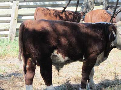
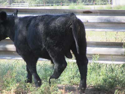
Bison – chronic hardware disease
This herd of bison was fed hay that contained chopped up cable. The herd experienced losses in the spring. The two bison in the image died in the fall from chronic disease. The hay was baled in a ditch to help winter the bison. It was a drought year and feed supplies were tight. A utility company had done line work along the ditch and some chunks of cable were left behind. The owner added magnets to the hay grinder when the problem was identified. Cattle producers often give rumen magnets, but bison will regurgitate magnets.
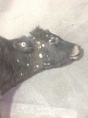
Calf – Streptococcus gallolyticus
Twelve 3 to 4 day old dairy calves became blind and died one day later. This calf had pus in the anterior chamber of the eye, a navel infection, arthritis, septicemia and meningitis. Poor calving area hygiene was likely related to the problem.
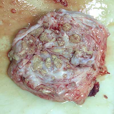
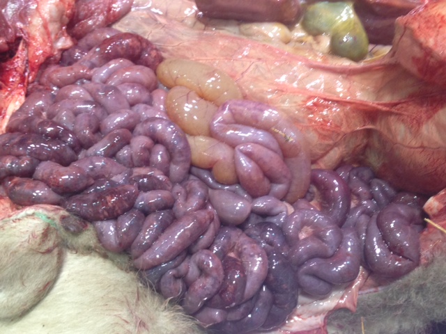
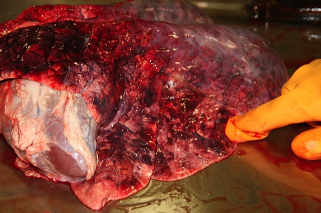
Sow – liver torsion
There was a history of sudden death. The death was caused by blood loss from a liver lobe torsion which then ruptured.
Calf – ventricular septal defect
This one month old beef calf was found dead. It had cardiomegaly and a 2.5-inch long high ventricular septal defect. The cause of death was heart failure
Bred heifer - Clostridium haemolyticum
Sudden death history. These heifers were pastured in an area of Minnesota known to have deer flukes. A wedge shaped area of necrotic liver was observed. The urine was red tinged. Clostridium haemolyticum was isolated from the affected liver. Parasiticides are available to help control flukes, and clostridial bacterins containing Clostridium haemolyticum should be used when needed.
Fat cattle – winter time hyperthermia
Twenty five of 359 cattle were found dead the morning after being delivered to a packing plant for slaughter. The cattle had red foamy nasal exudate. The cause of death was hyperthermia. The cattle were fat and covered with a thick winter hair coat. They went from an environmental temperature of -37F (-60F wind chill) to an indoor heated area where it was 70F. In addition, the floor was heated and bedded. Exhaust fans may also have malfunctioned. No evidence of infectious disease or toxicosis was found.
Lamb hepatitis
Dead six-day-old lamb
The liver contained multiple 1-2 cm areas of necrotizing hepatitis. Fusobacterium necrophorum were isolated. The infection most likely entered through the umbilicus.
Cow pyelonephritis
One dead six-year-old cow submitted with history of weight loss. There was severe bilateral pyelonephritis. Trueperella pyogenes was identified.
Cow metritis
A dead adult cow was submitted after calving five days previously. She was having some neurological signs shortly before death (a rabies test was negative). The cow had dead twin calves. There was a history of dystocia. Severe bilateral metritis was present. A mixture of bacteria was identified from the uterus including Trueperella pyogenes and E. coli.
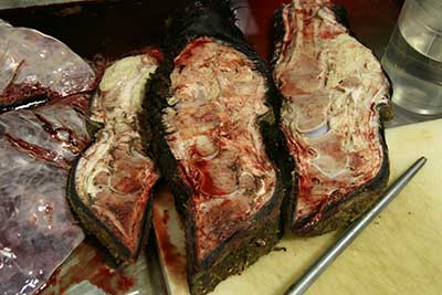
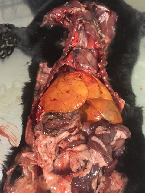
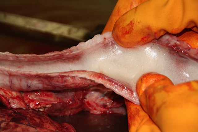
Hardware Disease
Hardware disease in beef cows is seen most often prior to calving when cows are being fed ground hay. The images show that the heart and one lung are stained with stomach contents that leaked along the penetrating wire. The growing near term fetus pushes the stomachs forward and precipitates hardware disease. It is important to keep wire out of hay bales. Hay grinding equipment should have functioning magnets to remove metal objects. Tire feeders should also be inspected for wear. Any tires with exposed metal should be disposed of.
Anaphylaxis
A dead six-week-old calf was brought to the laboratory. Two others from a group of seventy had died an hour after being given a booster vaccination. The lungs had marked pulmonary edema and trachea was filled with froth. No other significant findings were identified. A diagnosis of anaphylaxis was determined. The initial vaccination had been given at birth. Cattlemen should keep epinephrine available when giving booster shots or other injections. See your veterinarian to obtain this emergency treatment.
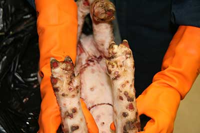
Pododermatitis in mink 1/27/16
A mink farm was experiencing ulcerative dermatitis on feet and other body areas. Arcanobacterium phocae was identified. This condition has been documented in Europe and Canada. This is the first confirmed case of this problem in the northern Great Plains.
Polyarthritis and tenosynovitis in a feeder calf 1/15/16
A feeder steer was brought to the laboratory for diagnosis of a lameness problem. Four of 200 head were affected by the problem. Both carpal joints and one tarsal joint were involved. Further examination of the affected legs found polyarthritis and tenosynovitis. Mycoplasma bovis and Trueperella pyogenes were identified from affected areas.
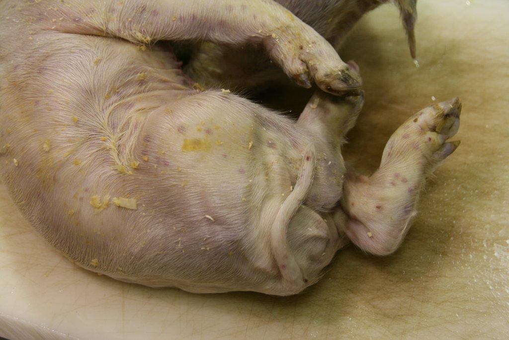
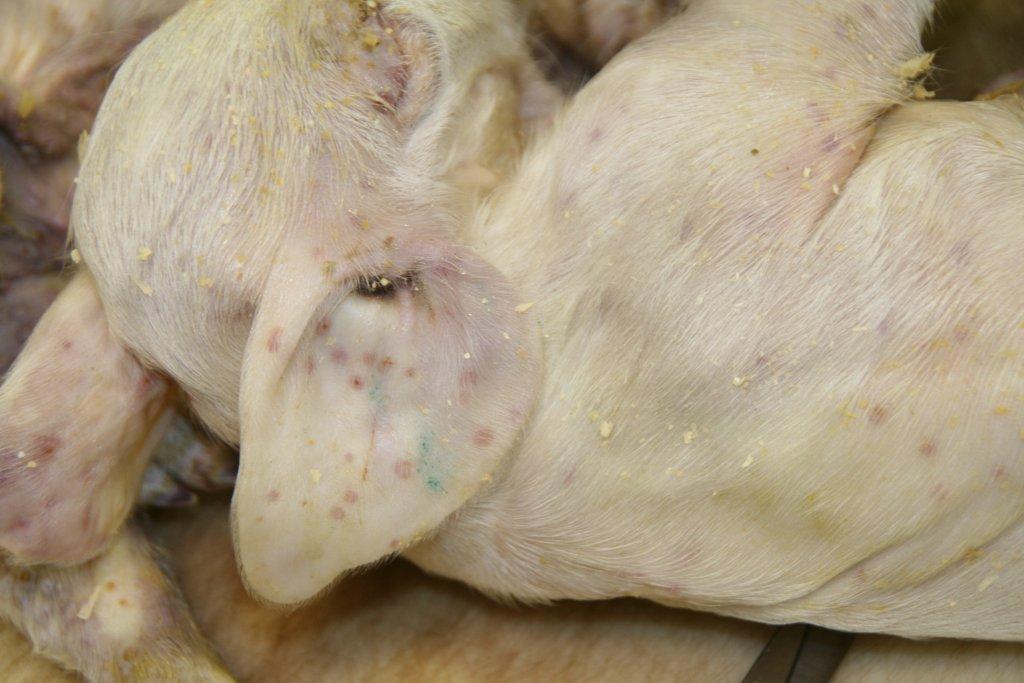
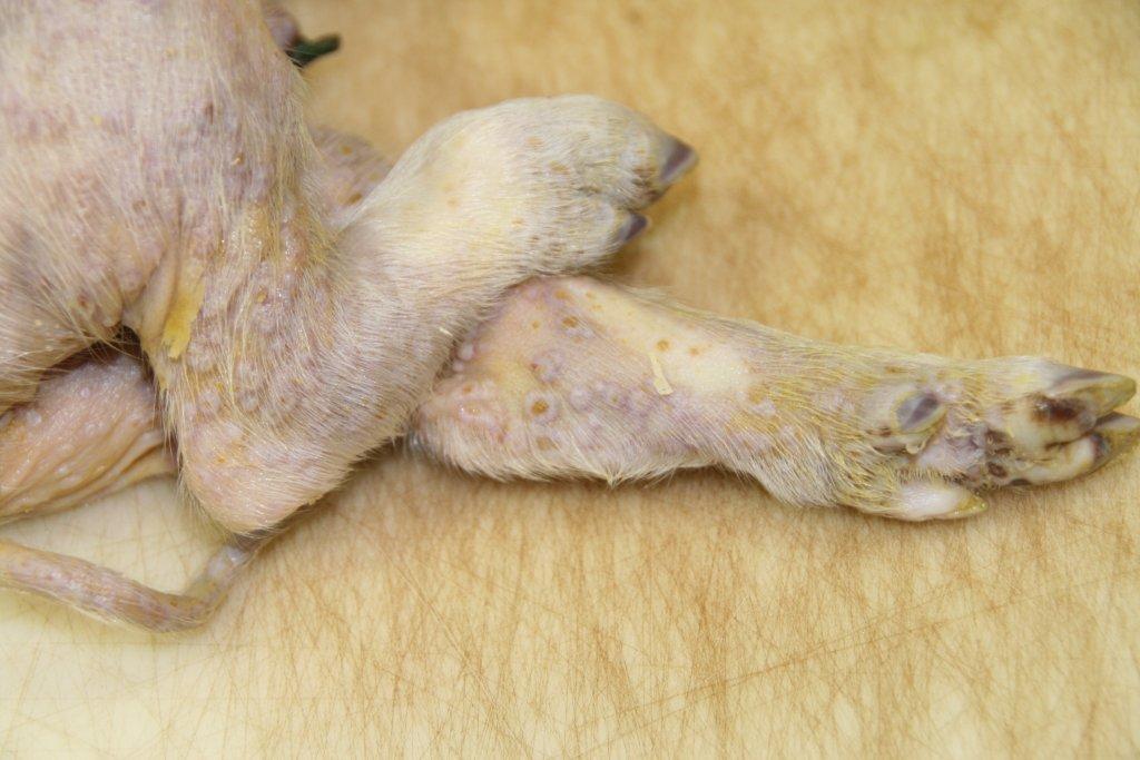
Swinepox 1/6/16
Low numbers of nursery pigs were experiencing skin lesions. The farm wanted to rule out FMD and Seneca Valley Virus. Swinepox was confirmed by PCR and sequencing.
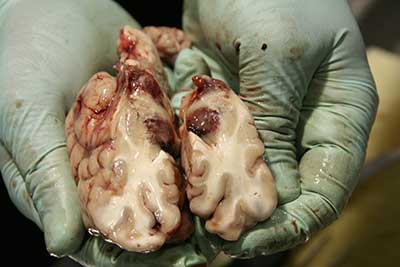
Histophilus somni meningoencephalitis in a weaned beef calf 10/29/15
This steer calf was recumbant in the morning and died later the same day. The brain had gross lesions of vascular thrombosis (thromboembolic meningoencephalitis). Histophilus somni was identified in the affected brain. Image 1; Image 2
Hepatic lipidosis in mink 10/29/15
Seven dead mink were submitted for examination. Hepatic lipidosis was the common finding. Stresses that result in several days inappetence during the fall period of furring up may lead to losses from this condition.
Ringworm in a steer 4/22/15
Ringworm is not an unusual cause of dermatitis in calves. Here is an image of a steer with ringworm (Trichophyton verrucosum).
Addison's Disease in a dog (hypoadrenocorticism) 4/21/15
A 5.5 year old castrated male Labrador retriever developed loss of appetite and lethargy on a Friday. He declined further over the weekend. A complete blood count and chemistry panel were conducted the following Monday. The results were suggestive of Addison's Disease (↑potassium, ↓ sodium, ↑ blood urea nitrogen, ↑ creatinine and Na/K ratio ~ 18). The dog did not respond to an ACTH response (stimulation) test which confirmed Addison's Disease. Treatment consisted of fluids, corticosteroids and desoxycorticosterone pivalate (DOCP). There was dramatic response to the treatment.
Bovine Virus Diarrhea in a steer 1/2/15
An eight month old steer died after a short illness with clinical signs of diarrhea and pneumonia. There were oral and esophageal ulcers. Peyer's patches in the ileum were hemorrhagic in appearance. A BVDV antigen ear notch ELISA test was positive. Fluorescent antibody tests for BVDV were positive on oral mucosa, esophagus and Peyer's patch from the ileum.
Aleutian Disease in Mink 11/10/14
A mink farm was losing mink with swollen image 1). Aleutian Disease is caused by a parvovirus. There is no vaccination or treatment. The diagnosis was confirmed with positive PCR (polymerase chain reaction) results along with the typical microscopic kidney lesions.
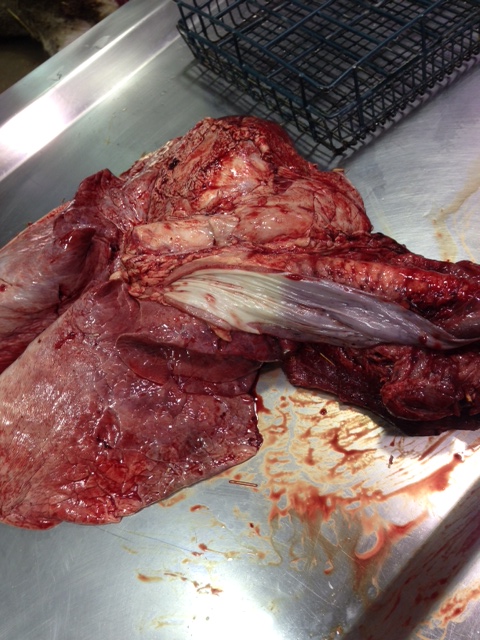
Mycoplasma bovis Infection in Domesticated Bison 9/08/14
Mycoplasma bovis can cause serious losses in domestic bison operations. Several South Dakota operations have been affected. The bacterial infection may cause pneumonia, arthritis, abortion and pharyngitis. See lung tissue demonstrating pneumonia.
Tail Necrosis in Beef Calves Associated with Ergot 8/26/14
A group of spring beef calves was losing tail switches and tail tips in August (image 1, image 2, image 3). Their creep feed contained significant levels of ergot (creep feeder). Read more about ergot and associated problems.
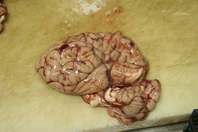
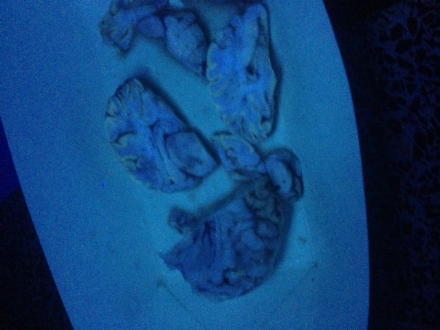
Polioencephalomalacia in a Calf 7/02/14
The brain from a 300 pound Holstein bull calf was submitted for rabies. The calf was recumbant, had nystagmus, paddling and muscle tremors. The rabies test was negative. Histopathology exams revealed cerebrocortical edema and necrosis. A diagnosis of polioencephalomalacia was made due to the typical brain lesions. Ultraviolet illumination of the formalin fixed brain demonstrated fluorescence of affected cerebral cortex (note apple green fluorescence of affected brain).
Paratuberculosis Vaccine Granuloma in a Dairy Heifer 6/13/14
This softball sized granuloma was in the subcutaneous breast tissue of the heifer. It was attributed to previous paratuberculosis vaccination. All tests were negative for Mycobacterium bovis.
Gastric Rupture in a Gelding 6/02/14
An eleven month old horse was castrated. The horse developed severe colic and shock. It was euthanized the following day. Necropsy examination revealed a gastric rupture.
Feline Tularemia 5/23/14
A young adult male cat was submitted for rabies testing. The rabies examination was negative, but and splenitis were found. Tularemia was confirmed by bacterial cultures on the spleen. Francisella tularensis organisms were identified.
Swine Pox 5/23/14
Two 12-15 day old pigs were submitted with dermatitis. Typical gross (image 1, image 2, image 3, image 4) and microscopic skin lesions of swine pox were observed. Electron microscopy exams confirmed the presence of poxvirus.
Mycoplasma bovis Pneumonia in a White-tailed Doe 5/07/14
A white-tailed doe was submitted after several unsuccessful treatments for pneumonia. The deer was from a commercial deer farm. The right lung demonstrated severe pleuropneumonia. The sectioned lung had diffuse caseous pneumonia. Mycoplasma bovis infection was confirmed with PCR examinations.
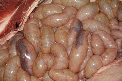
Clostridium perfringens type C Enteritis in a Beef Calf 4-17-14
A dead one and a half day old beef calf was brought to the laboratory after a brief history of bloody diarrhea. Severe hemorrhagic enteritis was observed. Clostridium perfringens type C infection was confirmed with bacteriology and PCR techniques. Another image of the severe enteritis. Necrotic enteritis was observed microscopically, and tissue gram stains show many large gram positive bacteria colonizing the necrotic intestinal epithelium.
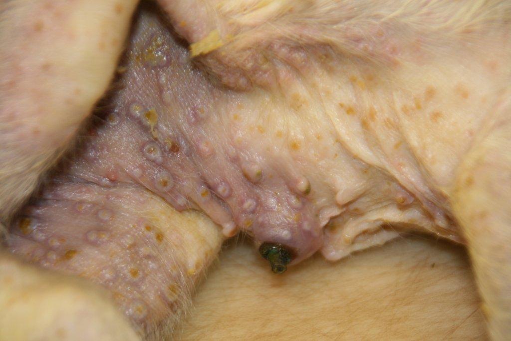
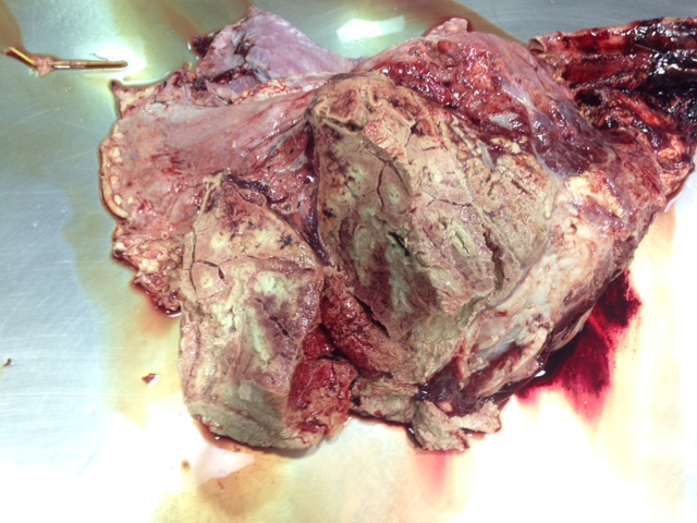
Erysipelothrix rhusiopathiae Endocarditis in a Pig 3-19-2014
Heart from a pot belly pig was received for examination. There was obvious vegatative endocarditis.
Erysipelothrix rhusiopathiae was identified on bacterial culture. This pig was one of five on the premise.
Streptococcus suis is found much more commonly in larger swine units.
Bloat in a Dairy Heifer 3-18-2014
A dairy heifer was found dead with it's head stuck in a gate. The carcass was bloated. Necropsy examination revealed a bloated rumen and an obvious "bloat line" in the esophagus. The cause of death was most likely suffocation due to bloat.
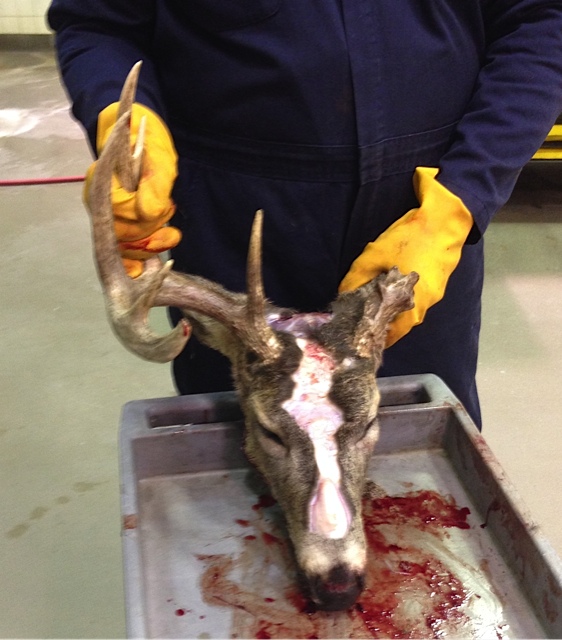
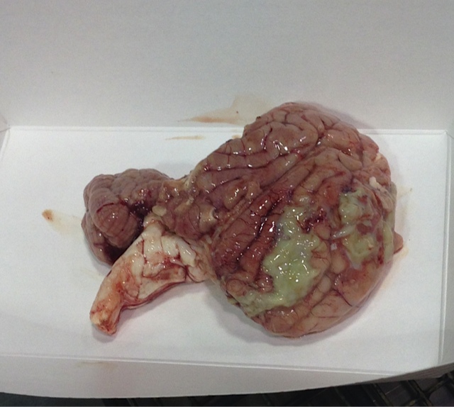
Meningitis in a White-tailed Buck Deer 3-07-2014
A white-tailed buck was submitted to determine the cause of unusual behavior. The deer was euthanized after demonstrating lethargy, circling and loss of fear of humans. The left antler was broken. Severe meningitis was observed at necropsy (notice pus on brain surface). Bacteriology exams identified Trueperella pyogenes (formerly known as Arcanobacterium pyogenes) from affected brain. Antler wounds from fighting may lead to this condition when they become infected.
Bovine Anaphylaxis Suspects 3-04-2014
Two one month old calves were found dead 3.5 hours after receiving a bacterin. Necropsy examination revealed froth coming from the nasal passages and in the tracheas. The lungs were edematous and mottled. Microscopic examination of lungs revealed pulmonary congestion and edema. Some interlobular lymphatics contained hemorrhage. A diagnosis of anaphylaxis was likely.
Holstein Calves with Pinkeye
A backgrounding operation was plagued by an extended pinkeye outbreak. Ocular swabs contained Moraxella bovis, Branhamella ovis and Manheimia (Pasteurella) haemolytica. Various ophthalmic treatments, oral and injectable antibiotics and eye patches didn't seem to solve the problems. The submitting veterinarian reported the outbreak halted within a couple days following ML V IBR vaccination and injectable vitamin A administration.
Group image.
Early clinical case.
Close-up view of early case - notice excess lacrimation and inflammation of the eyelids (blepharitis).
Chronic pinkeye case - notice corneal scar and excess lacrimation.
Tail Tip Necrosis and Polyarthritis
A live feeder steer in sternal recumbancy was brought to the laboratory. It had tail tip necrosis and polyarthritis. Arcanobacterium pyogenes were isolated from multiple joints and from lung abscesses.
Johne's Disease in a Beef Cow
This cow was losing weight and had diarrhea. The colon contained fluid contents. The ileal mucosa was thickened and the mesenteric lymph nodes were slightly enlarged. The Johne's DNA probe test was positive on the intestine.
Porcine Coccidiosis
One 16-day-old pig was submitted. Necropsy revealed fluid yellow diarrhea, a milk filled stomach and partly filled lacteals. Hostopath exams revealed atrophic enteritis with numberous coccidia in gut epithelium.
Canine Herpesvirus
One dead puppy was submitted for exam. Four puppies from two litters had died. Seven litters from this kennel were housed in the same building. Affected puppies were dehydrated, had open mouthed breathing, and cried before dying. Necropsy lesions included multifocal hemorrhages in kidneys and mottled lungs. Histopath exams revealed multifocal necrosis in liver, kidney and lung. Fluorescent antibody tests were positive on kidney for canine herpesvirus.
Coccidiosis in a Calf
A one-month-old beef calf was found dead in the pasture. Eyes were sunken and there were dark feces on the tail and hindquarters. The spiral colon contained blood -tinged contents and some shreds of fibrin. (Intestinal contents from the large intestine.) Moderate numbers of Eimeria zuernii were observed in the stool.
Lead Poisoning
A month-old beef calf died suddenly with no history of illness. Fifty cow/calf pairs were kept in a pasture with a tree grove. No significant gross lesions were seen at necropsy. Kidney was analyzed for lead and 86 ppm were found confirming lead poisoning. We see most cases of lead poisoning in calves in the spring. Calves are curious and seem to seek out lead sources which are often broken batteries.
Blackleg
Three of 180 six-month-old Holstein heifers died acutely with swollen necks. Necrotizing Myositis was found in the neck and necrotizing myocarditis was observed in the heart. Clostridium chauvoei was identified from heart and skeletal muscle. The heifers had not not received a clostridial vaccine upon arrival.
Lamb Pneumonia
Pasteurella haemolytica and Mycoplasma ssp. were isolated from the lungs of these four to six-week-old lambs. Lambs. Lungs.
Congenital Goiter in a Lamb
This flock did not receive iodized salt. 75% of lambs died shortly after birth. Swollen thyroid glands. Notice the enlarged thyroid gland next to a normal one from a different flock.
Pneumonia & Mastitis in a Yearling Feedlot Heifer
The feedlot operators reported coughing, lameness and diarrhea in a live heifer that was brought to the laboratory. They said she had been treated previously for pneumonia and was "bagging up". Pasteurella multocida, Haemophilus somnus and Actinomyces pyogenes were identified from lung. Actinomyces pyogenes was also identified from mammary gland. Mycooplasma bovis organisms were also identified from both lung and mammary gland.
Hardware Disease in a Feedlot Steer
Feedlot operators reported this steer was depressed, anorexic and reluctant to move prior to euthanasia. Hardware disease was found (endocarditis). Nephritis and encephalitis were also observed. Another recent hardware case. Actinomyces pyogenes was found in kidney.
Saddle Thrombus in a Calf
Owners reported that this 15-day-old calf suddenly started to knuckle over in both fetlocks. Skin on affected limbs was cold. The calf was euthanized. A saddle thrombus (aortic and iliac artery thrombosis) was discovered.
Congenital Hydrocephalus & Cerebellar Hypoplasia in a Calf
A live recumbent two-day-old calf was submitted for examination. Two other calves had similar signs. None were able to arise or nurse. Brain demonstrated severe gydrocephalus of lateral ventricles and severe cerebellar hypoplasia.
Ergotism
Two lame 10-month-old Holstein steers were brought to the ADRDL for examination. They were part of a mixed group of forty 300-500 lb. calves. The owner reported that 30 out of 40 animals were affected and that some animal had sloughed feet. The hind legs had a clear line of demarcation above the dewclaws (Image 1, Image 2). The skin below the lines was cold and hard. Microscopic examination of affected skin revealed full thickness necrosis of epidermis with underlying thrombosed vessels in the dermis. A ration consisting of corn, oats and pellets had been fed for about a month prior to the start of the problem. The oats contained many ergot bodies. Chemical analysis of oats confirmed a very high level of ergopeptine alkaloids.
Salt Toxicity
One dead adult beef cow was brought to the ADRDL for examination. She was one of four sudden cow deaths in a group of 60 cows and their calves a couple of days after a late fall blizzard. The cows had been in a grass pasture during the blizzard and were brought home to process the calves. Access to water was limited during the storm. Several bales of hay were fed to the cows. No significant gross lesions were observed in the cow. Further discussion with the submitting veterinarian revealed the fact that the cows had eagerly consumed salt when they arrived at the home farm. Chemical analysis of brain tissue revealed 2,050 ppm sodium. These results confirmed salt toxicity/water deprivation. Levels above 1,800 ppm in cerebral tissue are sufficient to make a tentative diagnosis. No further losses were reported.
Listeriosis
Twelve abortions occurred and two cows died following the opening of a new bag of silage. The animals were from a group of 68 head. Listeria monocytogenes was isolated from cow and fetal tissues. Cultures were negative on silage. Losses stopped after the feed was changed and the cows were supplemented with oral oxytetracycline. A "hot spot" of L. monocytogenes contaminated silage was suspected as the cause of the problem. Bovine abortion samples were discussed in the December 1998 Animal Health Matters Newsletter.
Toe Abscesses
A live 500 pound heifer was submitted from a group of feedlot heifers. The animals had been purchased three weeks before at a salebarn. The heifer had a history of rear limb lameness and swollen feet. The problem did not respond to antibiotic treatment, and the lameness progressed to recumbancy. Three other animals at the feedlot were down with a similar history. The toe tips were abraded and oozed foul smelling, black fluid when squeezed. Lateral claws of the hind feet were split which revealed pedal osteitis at the tip of P3 and a cavitated area undermining the sole and hoof wall. A hock joint contained inspissated suppurative exudate. Lungs had multiple necrotic lobules, some with large cavitation. Bacterial cultures identified Actinomyces pyogenes and Bacteroides sp. from lung and joint swabs. Pasteurella multocida was also identified from lung and Mycoplasma arginini from joint. This syndrome is known as tow abscess and can occur in cattle handled on rough surfaces. The claw tips are worn until there is white line separation which then allows penetration of dirt and manure into the claw. Bacterial infection with various aerobic and anaerobic microbes results in pedal osteitis, ascending foot infection, and bacteremia. The problem must be identified as soon as possible for treatment to be successful. The interdigital space is usually not affected, as in footrot. Recommended treatment is trimming the toe to allow drainage (excessive trimming will cause increased lameness) and broad spectrum antibiotic therapy. Some veterinarians will apply a block to the unaffected claw.
Copper Deficiency in Goats
To to three-month-old kids (goats) went down on back end. Animals alert and still eat. They had three other kids the same thing. Most get weak in hindquarters first but can get weak in front first.
Gross Necropsy - No gross lesions, gastrointestinal tract full of normal content.
Microscopically - Pulmonary edema and congestion, brevity of Purkinje cells in the cerebellum and axonal degeneration in the peripheral nerfes (namely sciatic nerve).
Chemical Analysis - Liver copper was 1 ppm (adequate = 25–150 ppm).
Diagnosis - Copper deficiency/enzootic atazia
Congenital Bovine Goiter
Partial alopecia, swollen neck and thyroid enlargement and myxedema
Mink Ranch Abortions
A mink ranch was experiencing an increased incidence of abortions. The normal rate was around 1% and it had increased to about 3% of the litters. Salmonella dublin organisms were identified from fetal stomach contents. The source of infection is usually contaminated feed.